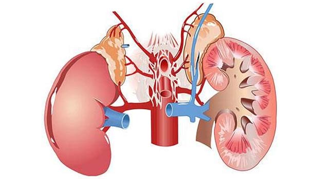
The kidneys are a complex organ of the urinary system that produces and stores urine. The renal pelvis serves as the reservoir for primary accumulation. Enlargement of the renal pelvis in a child is most often a congenital pathological process, less often acquired.
In childhood, in the vast majority of cases, an enlarged renal pelvis is a congenital pathology. Congenital causes are due to negative impact external and internal factors on the mother’s body in the first trimester of pregnancy, when the formation and development of the organs of the urinary system occurs. Under the negative influence, disturbances occur in the structure of the urinary organs. Vesicoureteral reflux and increased intrarenal pressure may develop.
Similar congenital anomalies occur in newborns if the mother was influenced during pregnancy negative factors environment: poor environment, stress, poor water quality. Also, expansion of the renal pelvis can occur under the influence of intrauterine infections and bad habits of the mother. Plays a big role genetic information- if cases of or have been diagnosed in the family, the likelihood of developing pathology increases.

In premature babies, the risk of pathology increases, which is caused by incomplete maturation internal organs and weakness of the abdominal wall.
Acquired enlargement of the renal pelvis in childhood is possible against the background of the development of infections, inflammations in the urinary organs or kidneys, as well as their injuries, oncology, etc. If the endocrine system is disrupted, hormonal imbalances, or metabolic processes are disrupted, the load on the kidney and its vessels may increase, which leads to urine backflow.
Infants may experience a physiological enlargement of the pelvis up to 7 - 10 mm. The expansion of the renal pelvis can go through several stages.
A child with pyeloectasia feels well, without any signs of organ dysfunction. This continues until the infection of the urinary organs occurs, when the process of enlargement of the pelvis occurs rapidly.
If the renal pelvis is dilated, the child experiences symptoms of inflammatory processes in the urinary organs:
If the renal pelvis is dilated and inflammatory processes develop in it, an increase in the level of leukocytes and protein is detected in the urine of a newborn during laboratory tests. With the naked eye you can see sediment and flakes. At stage 2, an enlarged pelvis is accompanied by the appearance of blood in the urine. At stage 3, skin itching, convulsions, and clouding of consciousness occur.

In the case of an increase in the size of the collecting system and urolithiasis, colic occurs in the organs of primary accumulation of urine.
If a child has enlarged pelvis, this can lead to some complications, which is caused by impaired urine flow and stagnation. Most often, in childhood, inflammation of an enlarged organ develops together with pyeelectasis.
Most dangerous consequence There may be a rapid expansion of the renal pelvis due to their overflow, which leads to organ dysfunction. In medicine, this disease is called hydronephrosis.
If the renal pelvis is enlarged due to an abnormal structure of the ureter, renal reflux may develop. This pathology occurs when there is a small lumen of the ureter, when urine is thrown back during passage. The narrow lumen of the ureter, together with an increase in pressure in the bladder, leads to megaurethra - a rapid expansion of the ureter.
In boys with dilated pelvis, the ureter can immediately flow into the urinary canal, and in girls into the vagina. This anomaly is called ectopic ureter. Another complication in which the ureter is damaged is urethrocele. It is characterized as a disease in which the ureter at the point of contact with the bladder is inflated, and the lumen narrows.
The pathology is discovered accidentally during a routine ultrasound during pregnancy or during the first year of life. If the renal pelvis enlarges in a child, it is necessary to be observed by a pediatric urologist. During the first year of life, the baby must undergo a monthly urine test to assess the condition of the urinary system and timely diagnosis of possible abnormalities. Also, once every 3 months it is necessary to undergo a routine ultrasound examination to study the size and condition of the organ over time.
After a year, if the dynamics are positive, routine examinations are reduced. The child needs to have a urine sample every 2-3 months for a general clinical examination and an ultrasound scan every 6 months to assess the condition of the urinary system. As a rule, after 2 - 3 years, the number of examinations is reduced to 1 per year or carried out according to indications.
![]()
If a general clinical analysis of urine shows poor indicators (increased levels of leukocytes, protein, the presence of red blood cells), a bacteriological examination of the urine is required. Urine culture will allow you to determine the infectious or bacteriological pathogen, the presence of which has led to a deterioration in urine parameters. Such a study is also necessary for choosing a method of therapy, as it allows one to determine the resistance of the infection to antibacterial drugs.
If there is a suspicion that the child’s renal pelvis is dilated due to stones or tumor formations, it is necessary to undergo X-ray diagnostics or computed tomography. These methods can give a clearer picture, unlike ultrasound, and allow you to see problems that are not visible on ultrasound images.
If there are no results from research using the above methods, endoscopic examination is used. This method allows, using an endoscope, to determine how dilated the renal pelvis is, to assess the condition, structural anomalies, and to determine obstacles in the way of urine discharge.

Minor enlargements during the first 2-3 years of life, in the absence of complications, require a wait-and-see approach. The child must undergo a routine ultrasound examination and submit urine for a general clinical analysis. Waiting tactics are applicable for congenital pathology, when the pyelocaliceal system is not fully formed.
Pathology requires treatment for abnormal structure of the urinary tract organs, the presence of stones, tumors, rapid growth of the collecting system, infection and other complications.
Children with an enlarged pelvis are prescribed medications in accordance with the identified complications. For pyelonephritis, it is necessary to undergo a course of antibacterial and anti-inflammatory therapy. Physiotherapeutic treatments can also be used.
For urolithiasis, medications are prescribed to dissolve and remove stones from the kidneys. At pain syndrome antispasmodics and painkillers are used. Drug therapy is used alone or in combination with surgical treatment.

Surgery is necessary for complications of pyelonephritis, large sizes stones, oncology, hydronephrosis, abnormal narrowing of the urinary tract. For dilated pelvis in childhood, the laparoscopic method is most often used, which is minimally invasive with short term postoperative recovery. Contraindications to laparoscopy include premature or low birth weight children, as well as those who have other developmental abnormalities.
If a child's pelvis is dilated, this is not a reason to panic. It is necessary to consult a doctor who will examine the child, determine the size of the organ, the degree of deviation from the norm and assess the general condition of the child. Based on these data, he will prescribe treatment or advise observation and waiting for the urinary system to fully mature.
IT IS IMPORTANT TO KNOW!
For the treatment of kidneys and urolithiasis, patients use the innovative development of Russian scientists, which has undergone clinical trials and proven its effectiveness. Renon Duo - just 3 capsules will eliminate lower back pain, kill bacteria and pathogenic flora, and effectively help with swelling!
The funnel-shaped cavity that serves to collect urine from the renal canals is called the renal pelvis. Such a cavity is located in each of the kidneys. It is due to its contraction that urine moves into the bladder through the ureters. If upon examination of the child an enlargement of the renal pelvis is revealed, there is no need to panic. Usually this condition is a normal physiological phenomenon that spontaneously resolves when the baby reaches the age of 1-2 years.
Usually, when the pelvis is dilated, you should not panic
This pathology most often occurs in male children; girls have an increase almost 4-5 times less often. In this case, a distinction is made between unilateral or bilateral pathology. When the renal calyces are dilated together with the pelvis, renal hydronephrotic transformation is observed in children. Enlargement of the kidneys and ureter is called megaureter.
Many parents who are faced with this problem are interested in what is the reason that the child’s renal pelvis is dilated? Here, first of all, factors such as genetic inheritance or the effect of toxic drugs on the fetus and the mother’s body should be considered.
As a rule, pyeloectasia (enlargement of the renal pelvis in newborns) develops when the outflow of urine is impaired. This occurs when the ureters are too narrow and unable to pass required quantity liquids. It accumulates in the pelvis, causing their deformation.
Also, the cause of the development of pathology in children can be ureteral reflux, in which the fluid that gets into the bladder is thrown back into the kidney. There is a congenital disorder of urine flow. Normally, the valve located in the vestibule of the bladder closes it tightly, preventing fluid from rising back up. If the valve does not work, the problem described above occurs.
In addition, increased pressure in the bladder can lead to pathology of the renal pelvis. This condition usually occurs in a child who has problems with the nerve supply to the genitourinary area.
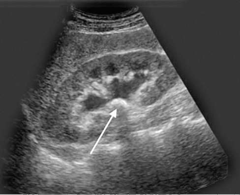
Regardless of what reason led to the expansion of the renal pelvis in a child, it is one of the congenital pathologies. The increase can be seen during ultrasound diagnostics as early as the 5th month of fetal development.
The size of the renal pelvis in a newborn should not exceed 10 mm. But even if pathology is observed, it does not cause any inconvenience to the baby, causing neither painful nor unpleasant sensations. That is why pathology is most often discovered by chance, when applying for an ultrasound examination for some other reason. If an enlarged pelvis is detected in the kidneys, the baby is prescribed an x-ray diagnosis of the bladder and kidneys.
Typically, pyelectasis that does not go away as the baby grows can lead to the following complications caused by impaired urine outflow:
As the child grows, mild forms of all of these diseases usually go away on their own. Treatment requires only severe pathology, in which even surgical interventions are possible.
If in the first days or months of life an enlargement of the renal pelvis is detected in children, it is necessary to constantly monitor changes in their size. This can be done using ultrasound diagnostics and test monitoring. In cases of mild kidney pathology in newborns, the doctor should prescribe an ultrasound scan at least once every 3 months.
The best way to monitor the kidneys is ultrasound
If the child's tests show the presence of inflammation, infection, or the size of the renal pelvis has increased sharply, a full urological examination is necessary, which includes methods such as radioisotope studies, intravenous urography and cystography.
Using these methods, the doctor will be able to form a complete picture of the disease, determine the degree of development of the pathology, and also detect or exclude hydronephrosis, ureterocele, etc. In most cases, the pathology is completely curable, and the child will “outgrow” it on his own upon reaching a certain age.
For severe pyelectasis in children, treatment mainly comes down to taking medicines, aimed at restoring the outflow of urine from the enlarged pelvis. If medications are not able to eliminate the problem, a decision is made to undergo surgery to remove the obstacle that prevents urine from entering the bladder.
Nowadays, modern medicine has technology in which such an operation is performed using a bloodless method, using an endoscope and tiny instruments inserted into the urinary tract of small children. Ureteral reflux and some other diseases are cured endoscopically.
If a child is diagnosed with pyelectasia, there is no need to despair: dilated pelvis and complications are successfully cured and do not have a malignant course. The main thing is to regularly visit a nephrologist and undergo the ultrasound examinations prescribed by him on time.
Congenital anomalies of the urinary system are increasingly encountered in pediatric practice. A common pathology is considered to be dilation of the renal pelvis in newborns, or, which is not a serious pathology, but still requires constant monitoring and supervision by a doctor. An enlarged renal pelvis in a newborn is diagnosed more often in boys and 5 times less often in girls. In mild forms of the pathology, the prognosis is favorable, but when the disease progresses, the functioning of the kidneys and urinary system is significantly impaired, which can lead to severe and sometimes irreversible processes. According to statistics, dilated pelvises are more often present on the left kidney; damage to the right organ or bilateral damage is less often diagnosed.
The renal pelvis in newborns is a cavity formation in which urine accumulates before it moves further into the ureters. If this function is disrupted, the renal pelvis enlarges and expands, urine begins to accumulate in the renal pelvis, its outflow is disrupted, and stagnant processes may appear.
It is possible to determine that the renal pelvis is dilated in a newborn in the prenatal period, when the woman undergoes screening ultrasounds. Good visualization of the kidneys is observed already at the 17th week of antenatal development. Normally, the size should not exceed 4-5 cm. For a newborn baby, the norm is 6-7 mm. If there are deviations, the doctor assesses the general condition of the baby, determines the cause, and, if necessary, prescribes treatment. In some children, the norm may be 8 mm, but if the diameter of the organ is from 8 to 10 mm, this condition is already considered a pathology and is called pyeloectasia.
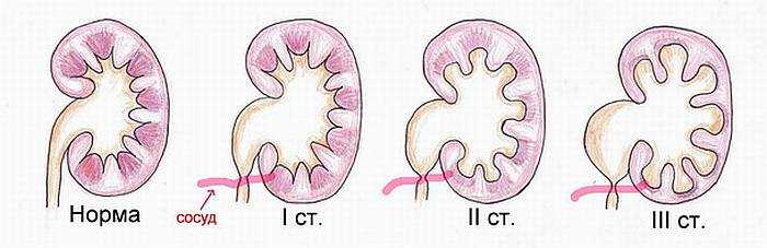
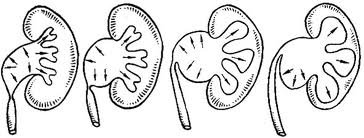
Stages of pyeelectasis
Enlargement of the renal pelvis in a newborn may appear at the most various reasons, but in 70% the pathology is hereditary. If one of the parents had such a disease, there is a high probability that the baby will have a pathologically dilated renal pelvis after birth. Other reasons that can trigger the appearance of this pathology include:
Provoking factors that can lead to an enlargement of the renal pelvis in a newborn include long-term use of certain medications by the mother during pregnancy, alcohol consumption, smoking, and a history of chronic diseases. In some cases, the pathology is detected after birth, manifests itself against the background of ionizing radiation, intoxication of the woman’s body, or during infectious diseases.

In pediatric nephrology, pyelectasis is classified as:
The most common pathology is the left kidney and extremely rarely the right or both. This condition is also classified according to its etiology:
Regardless of the classification of the disease, if the pelvis of a newborn is dilated, the doctor must prescribe a series of studies that will help determine not only the cause, but also the stage, and draw up rough plan for therapy.
As pyeloectasia progresses, it goes through several stages, each of which is accompanied by certain changes in the renal tissues and urinary system.
Kidney pelvis in a newborn at this stage increased slightly, do not interfere with the functioning of the organ. The child does not experience any unpleasant sensations, and the pathology itself can only be diagnosed using ultrasound during fetal development or immediately after birth.
The second stage of the pathology is accompanied by a pronounced expansion of the pelvis, there is damage to the external tissue of the organ, its function is reduced by 40%. At this stage of the disease, severe symptoms may be present, which forces parents to consult a doctor. The child becomes restless, often cries when urinating, and there may be blood in the urine.
The most severe stage of the disease, which is characterized by severe symptoms. The child has an enlarged pelvis and the kidney itself, significantly reduced urine production, increased body temperature, pain when urinating and other symptoms that require a medical examination. The kidney tissue is significantly damaged, and when the pelvis is greatly expanded, it puts pressure on other tissues.

The likelihood of complications with pyeloectasia in a newborn is low, but still in some cases, as the pathology progresses, the following symptoms may appear:
In order to reduce the risk of complications, you need to recognize the disease in time and carry out the necessary treatment prescribed by your doctor.
You can recognize an enlarged renal pelvis in a newborn using ultrasound, which is the most informative and safe for the baby and allows you to recognize the dilation of the urinary ducts, assess the size and function of the urinary system. The doctor may prescribe additional research methods, including: laboratory research, cystography, x-ray, the results of which will allow you to get a complete picture of the disease, select optimal scheme treatment.

If the newborn has a mild form of the disease, treatment is not carried out. The child is registered with a doctor and must undergo regular ultrasound scans to monitor the condition. Very often, the pathology disappears on its own before the age of three. If the pathology is diagnosed during fetal development, the expectant mother should remain in the hospital under medical supervision until the birth of the baby. For such women, the number of ultrasounds required is increased to two monthly. This research method will allow you to monitor the functioning of the fetus’s kidneys and assess its general condition.
In cases where the pathology rapidly progresses or the second stage is diagnosed, the child is prescribed conservative treatment- taking medications that improve urine flow, as well as physiotherapy courses, regular ultrasound monitoring. Treatment may last several months. If the dynamics are positive, surgical intervention is not performed. If conservative therapy does not bring the desired results or the function of the urinary system is significantly impaired, the only treatment method will be surgery. The most commonly used method is laparoscopic or endoscopy, which are gentle procedures. The prognosis after surgery is favorable, the main thing is to recognize the disease in time and prevent its complications.


