Pyeelectasis of the kidneys is a pathology accompanied by an increase in the cavity of the pelvis. It can never be an independent disease. It is considered as an indirect symptom of impaired urine outflow from the pelvis due to the presence of infection or structural abnormalities of the filtering organs. Treatment should begin with eliminating the causes of renal pyeelectasis, otherwise the pathology will progress.
Renal pyeloectasis is divided into three degrees according to severity:
 The severity of the pathological process is determined by the preservation of renal function. It can also be established taking into account the average frequency of development of diseases of the urinary system, which are inflammatory and infectious in nature. Pathology can affect not only the renal pelvis, but also the renal pyelocaliceal system and ureters. They also begin to expand.
According to the extent of the damage, they are distinguished:
With bilateral pathology, two organs are involved in the process at once. In unilateral cases, pyeloectasia of the left or right kidney is observed. Pathology is more common in males. Moreover, pyeloectasia of the right kidney is most often observed due to its anatomical location. But the pathological process may include dilation of the ureters and calyces.
If this happens to the right organ, then a diagnosis of “pyelocalicoectasia of the right kidney” is made.
Depending on the cause that caused the pathological expansion of the renal pelvis, 4 forms of pyelectasis are distinguished:
In adults, the pathology most often develops when the ureter is blocked by a stone or a clot of mucus or pus, which forms during inflammatory diseases of the kidneys.
The pelvis dilates when the kidney wanders or prolapses as a result of twisting or bending of the ureter. In an adult, the pathological process can begin even when taking very large quantity water when the urinary system can no longer cope with the imposed load.
In the elderly bedridden people expansion of the pelvis occurs due to deterioration of ureteral peristalsis.
Renal pyelectasis is characterized by accompanying pathologies:
In adults, pyelectasis usually occurs without symptoms. But sometimes you can detect symptoms of the underlying disease, due to which the pathological expansion of the pelvis began. In this case, the urine begins to stagnate, which causes the development of kidney sclerosis. This name is given to necrosis of the tissue that produces urine. Necrosis is accompanied by renal failure, atrophy of the tissue of the filtering organs and pyelonephritis.
Often, dilation of the pelvis in a congenital form of pathology is detected in the fetus during a woman’s pregnancy, but sometimes in a newborn during the first ultrasound. It is noteworthy that this problem occurs up to 5 times more often in boys than in girls. Usually it has a congenital form. Less commonly, pathology develops against the background of increased growth of the body and its organs inside.
In adults, dilation of the pelvis most often occurs as a result of blockage of the ureter with a calculus. This condition is detected by performing urography, ultrasound or computed tomography of the kidneys (CT). To prevent the development of pyelectasis in patients urolithiasis It is recommended to carry out ultrasound examinations regularly. It helps to determine the static size of the pelvis before and after urination. In addition to ultrasound, cystography may be required to diagnose the pathological process.
Treatment of pyelectasis is determined after establishing the causes of its development. If the root cause is not established, the pathology will continue to progress, which will certainly lead to various kinds of complications.
Treatment of pyeelectasis in pregnant women is not required, as it goes away on its own after childbirth.
If there is an abnormal development of the urinary tract, treatment requires plastic surgery to correct it. But if there is a stone in the ureter, it must be removed surgical method. Thus, conservative treatment Pyelectasia of the renal pelvis does not exist. However, to stop the progression of the pathology, it is necessary to prevent stagnation of urine. To do this, empty your bladder more often. Treatment includes mandatory use of uroantiseptics that prevent inflammatory and infectious diseases urinary system.
Good Nurse
Have you found necrosis? Complicated by pyelonephritis xD
Good Nurse
Not “Necrosis is accompanied by pyelonephritis,” but “Pyelonephritis may be complicated by necrosis.” Think about what you write! Necrosis is tissue death, and pyelonephritis is inflammation. Necrosis and sclerosis are also different things. Sclerosis is by no means tissue death. This transformation of parenchymal into connective tissue leads to the impossibility of blood filtration! I thought I had stumbled upon a normal article, but now I VERY doubt it!
Pathology characterized by anatomical enlargement renal pelvis, called renal pyelectasis. The pelvis is a place where urine from the kidneys accumulates, which subsequently goes to the ureters. Pyeloctasia of the kidneys is not an independent disease; pathology indicates disturbances in the functioning of organs that are involved in the outflow of urine.
Why does the renal pelvis become enlarged in adults? The renal calyx accumulates and processes fluid entering the body, then it enters the pelvis, where it turns into urine. Due to certain processes, urine cannot move in full quantity into the ureter, which is why the renal pelvis becomes distended (normally slit-like). The condition rarely goes away on its own. Enlargement of the pelvis is usually divided into the following degrees:
The pathology is divided, based on what factors provoked the expansion of the renal pelvis, into the following types:

The dilated renal pelvis is divided depending on the degree of damage to the sides:
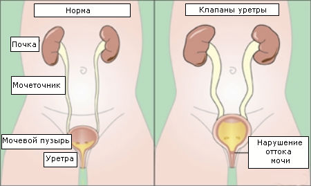 Urethral valves are a congenital pathology of the mucous membrane of the urethra.
 The following causes of pyelectasis are distinguished:
 The disease has no characteristic symptoms, so if there is the slightest malfunction in the urinary system, you need to undergo diagnostics.
 Enlargements of the renal pelvis occur without their own symptoms. More often, the pathology does not make itself felt for a long period and does not cause any inconvenience. Pyeelectasis in adults is in most cases diagnosed during examinations carried out to determine other diseases. The following symptoms are observed with pyelectasis:
 In most cases, boys are more prone to the occurrence of pathology.
 Experts are convinced that moderate pyeloectasia of the right kidney is more often observed in children than pyeloectasia of both kidneys and pyeloectasia on the left. Often the pathology is diagnosed in male children. If we talk about newborns, then pyeloectasia in them often represents congenital pathology and is caused by abnormalities in the structure of the ureter and other organs of the urinary system. It often happens that the pathology goes away on its own before the age of 2, however, if pyelectasia does not go away after growing up, the child should be systematically taken for an ultrasound examination, which shows an echo picture of the enlargements.
Factors influencing the development of pyelectasis in children:
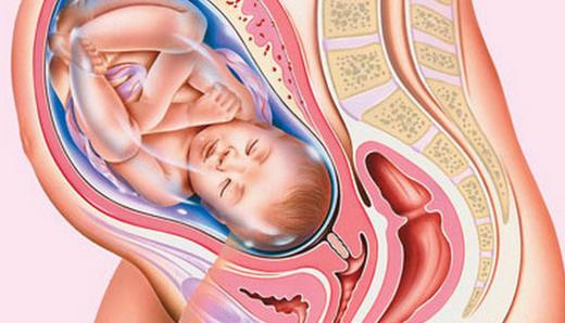 In pregnant women, pressure on the ureter causes an enlarged uterus.
 The condition when the renal pelvis is dilated in pregnant women is provoked by pressure on the ureter from the enlarged uterus (the renal calyces may also be affected). However, this is not the only reason; pyeelectasis can also develop due to hormonal imbalances. Pyeelectasis of the left kidney is diagnosed during pregnancy several times less often than the right one. The pathology is called “passing” because it can disappear on its own without the use of medical manipulation. This happens after a woman gives birth.
It should be noted that when diagnosing pyeloectasia during pregnancy, it is important to accurately establish whether the anomaly has developed due to position or whether it has begun somewhat before pregnancy. In case of pathology, they do not resort to termination of pregnancy, however, if pyeloectasia is chronic, this can seriously affect future births. Thanks to this factor, the permissibility of pregnancy in chronic pathology can be determined only after a proper examination has been carried out and the condition of the kidney has been studied.
Kidney pyeloectasia in adults is dangerous due to the factors that provoke it. Impaired release of urine from the kidneys, if not treated in a timely manner, provokes compression and then atrophy of the organ tissue. Because of this, the kidney begins to function worse over time, which often leads to its complete failure. Pathology can provoke the development of chronic and acute pyelonephritis (inflammation of the kidneys and calyces), which has a detrimental effect on the organ. That is why, if you suspect pyeloectasis, you should not delay contacting a doctor and undergo all the necessary tests in order to find out exactly what is to blame for the appearance of dilated pelvis, you need to undergo an examination and begin to treat the problem as soon as possible.
 The volume of the renal pelvis can be studied using ultrasound diagnostics.
 Conditions when the pelvis is enlarged in an adult are determined by ultrasound, during which specialists study the volume of the renal pelvis during and after the urination process. Additionally, the echo pattern and the significant size of the pelvis (the norm is 6 mm or more) and their changes over the next year, if any, are examined. When the size increases, it means that pyeelectasis is progressing. Then the patient will have to take general analysis urine. If the data obtained is insufficient, additional examination methods are used, including urography (an x-ray method for examining the urinary tract, which is based on the ability of the kidney to secrete certain radiopaque substances previously introduced into the body) and cystography (an x-ray examination method, the purpose of which is to obtain an image of the urinary cavity by filling it with a contrast agent).
A pathological condition such as pyelectasia refers to the expansion of the renal pelvis. This disease is much more common in boys than in girls. Pathological changes can be minor (moderate pyelectasis) and quite extensive.
It is important! Kidney disease, pyeloectasia can affect one (right-sided and left-sided pyeloectasia) or both kidneys. Mild forms of pyelectasis most often disappear on their own, while severe forms almost always require surgical intervention.
What are the renal pelvises?
The renal pelvis is the name given to the special cavities in which urine collects. From the pelvis, urine enters the ureters, through which it goes directly to the bladder.
The appearance of pyeloectasia may be influenced by genetic predisposition, as well as harmful toxic effects on the body of the mother and fetus during gestation. In cases where there is an obstacle to the natural outflow of urine, urine accumulates above this obstacle, which is what leads to the expansion of the renal pelvis.
It is important! Moderate renal pyelectasis, in most cases, does not have a negative impact on the health of the unborn child. During pregnancy, a spontaneous disappearance of moderate dilation of the renal pelvis is observed.
The presence of severe pyelectasis (more than 10 mm) may indicate a progressive difficulty in the outflow of urine from the kidney. This pathological process is characterized by an increasing nature, leading to atrophy of the kidney tissue, compression and decrease in kidney function.
It should be noted that a violation of the outflow of urine is often supplemented by the addition of pyelonephritis (inflammation of the kidney), this significantly worsens the state of health.
Is it necessary to examine a child after birth for the presence of pyelectasis? Moderate pyeelectasis of the kidney in most children spontaneously disappears against the background of growth of the organs of the urinary system.
For moderate pyeloectasia, it is quite sufficient to conduct regular ultrasound scans every 3 months after the birth of the baby. 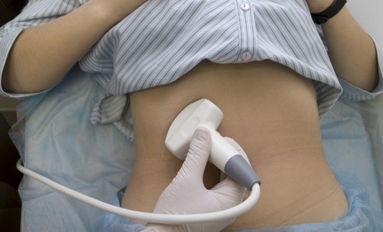
When a bacterial infection occurs, the use of antibiotics is necessary.
As the degree of pyelectasis progresses, a more detailed urological examination is indicated.
There is also surgery to remove the obstruction to the flow of urine. Certain surgical interventions are successfully performed using endoscopic methods - without open surgery, miniature instruments are used that are inserted directly through the urethra.
During an ultrasound, a characteristic change in the size of the pelvis is observed (before and after urination):
Information about possible complications
Treatment tactics are determined exclusively by a specialist - a urologist; the selection of treatment is based on the results of the studies. U
It is important! The main task is to eliminate the underlying cause of the disease.
Particular attention should be paid to the prevention of pyeloectasia - compliance with instructions during pregnancy, and limiting fluid intake, as well as complete and timely treatment of related diseases. After all, it is much easier to prevent the development of a pathological process than to then struggle for a long time and painfully with various complications.
Kidney pathology is not just words; behind them lie destroyed hopes, crippled destinies and the inability to live a full life. That is why, considering this problem, I would like to believe that people will need the information only for informational purposes.
This article will discuss bilateral renal pyelectasis. This is a condition in which there is a picture of pathological expansion of the renal pelvis. This diagnosis cannot be considered an independent disease; rather, it is a sign of impaired outflow of urinary fluid directly from the pelvis. The root cause of such a failure can be any anomaly in the structure of the paired organ or a previous infection.
The danger of this anomaly, namely the difficulties associated with urine discharge, can cause atrophy of the renal tissue, which will significantly affect the decrease in the functionality of the kidney. In the future, inflammatory processes may occur, leading to sclerosis of the excretory system organ.
Unfortunately, pyeelectasis of both kidneys is characterized by a severe and protracted course, and frequent relapses are also possible.
Organic acquired causes of the development of pathology are:
Organic congenital are pathologies that manifest themselves at the stage of formation of the walls of the upper urinary tract, kidneys or ureter.
Dynamically acquired reasons for the development of the anomaly are:
Dynamic congenital ones are phimosis, neurogenic disorders during urination, and narrowing of the urethra.
In adulthood, this pathology can be asymptomatic. Most likely, the person will experience symptoms of the disease, which became the main cause of the development of the anomaly.
Such a violation is very often a companion to such ailments as:
This type of anomaly is easy to notice during intrauterine development of the fetus, as well as in the first year of the baby’s life. This pathology can also occur during intense and sudden growth. internal organs. In adulthood, such an unpleasant pathology is most often caused by stones.
As for treatment, it is prescribed only by a doctor, based on preliminary tests. The main method of treatment is to eliminate the provoking disease. If the anomalies are congenital, then it is impossible to do without surgical intervention.
Renal pyeloectasia is a pathology accompanied by dilation of the renal pelvis and difficulty in the outflow of urine. The disease occurs in both children and adults and is often combined with other changes in the urinary system. Enlargement of the pelvis and ureter is not an independent disease and occurs in various pathological processes.
Before finding out the causes and starting to treat the disease, you should understand the structure of the kidney. This paired organ is located behind the peritoneum and is covered with a capsule that protects from various damage, inside which there is a urine storage system consisting of many small cups. The latter, connecting with each other, form a pelvis. The urine processed by the kidney then enters the ureter and is excreted from the body through the bladder and urethra.
Enlargement of the renal collecting system is called pyeloectasia. Quite often this phenomenon is accompanied by a change in the size of the ureter. This is not surprising, because the factors contributing to the formation of these conditions are usually the same. Why does renal pyeloectasia develop?
Possible causes of the disease:
All these reasons result in urine being retained in one area of the urinary system. Stagnation of urine inevitably provokes expansion of the pelvis and ureter. This phenomenon occurs both on the right and on the left. It is possible that bilateral kidney damage occurs simultaneously.
In newborns, dilation of the renal pelvis is usually a result of underdevelopment of muscles and ligaments. This condition is more common in premature babies and children born with low birth weight. In infants, renal pyeelectasis quite often accompanies perinatal lesions nervous system. With this pathology, muscle hypotonicity also develops, which leads to impaired urine excretion in the child and its stagnation in the kidneys.
You should know that kidney enlargement in newborns does not always require treatment. As the child grows, moderate uncomplicated pyelectasis goes away on its own. Therapy may be required only if the disease is accompanied by severe impairment of renal function and the development of inflammatory processes.
It has been noted that pyeloectasia of the right kidney in children is somewhat more common than enlargement of the pelvis on the left. Mostly boys are affected. In newborns, the pathology is usually congenital. Often, pyeloectasia in infants is combined with malformations of the ureter and other genitourinary organs.
Pyeelectasis does not have its own specific symptoms. This pathology may not manifest itself for a long time and not cause concern to the child. Often, dilation of the pelvis is first detected in adults by chance during examination for another disease. Prolonged course of the disease leads to the development of pyelonephritis - inflammation of the kidneys.
With congenital pyelectasia, pyelonephritis manifests itself in childhood or adolescence. The child’s body temperature rises, urination becomes more frequent, and lower back pain appears. Over time, stagnation of urine leads to kidney atrophy (reduction in its size) and sclerosis (growth of little functional connective tissue). In adults, the disease can cause kidney failure.
The severity of the symptoms will depend on the severity of the disease. If only the left or right kidney is affected, the child may not have any symptoms for a long time. A healthy kidney takes over all the functions of the affected organ and ensures normal urine excretion. Bilateral pyeloectasia reduces the performance of organs much more quickly and forms renal failure.
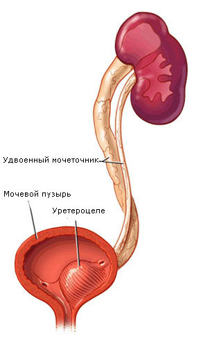
Congenital pyeelectasia is often combined with other pathologies of the urinary system:
 Kidney pyeloectasia in children is clearly determined by ultrasound examination. Already in the first trimester of pregnancy, the doctor can detect dilation of the pelvis and ureters in the fetus. In newborns, the disease is often detected by chance during routine medical examinations. If pyelectasis is detected in a child, an X-ray contrast study (urography) is performed to assess kidney function.
Treatment methods for pyelectasis depend on the severity of renal dysfunction. If the child's urine output is not severely impaired, doctors prefer a wait-and-see approach. Pyeelectasis in most newborns goes away on its own without treatment. Regular ultrasound monitoring and specialist supervision is the best tactic for minor deformities of the ureter and moderate expansion of the pelvic system.
If the disease is complicated by pyelonephritis, specific therapy is prescribed. Treatment includes the use of antibacterial and anti-inflammatory drugs. Physiotherapy has a good effect, as well as therapeutic exercises that improve the flow of urine from the affected organ.
Surgery is performed to restore normal urine flow. To do this, correction of congenital malformations, tumors and other structures that interfere with the normal functioning of the kidneys is carried out. IN last years Surgeries on children and adults are performed using minimally invasive endoscopic techniques. After the procedure, most patients are able to live a normal life without restrictions.
Kidney pyeloectasia is a disease that modern medicine can cope with quite successfully. Timely treatment and compliance with the doctor’s recommendations will allow the child and adult to get rid of all manifestations of the disease and avoid the development of serious complications.


